Home » Without Label » Upper Back Muscles Diagram - Back Muscles Back Muscle Diagram Muscleblitz Com / Dummies has always stood for taking on complex concepts and making them easy to understand.
Upper Back Muscles Diagram - Back Muscles Back Muscle Diagram Muscleblitz Com / Dummies has always stood for taking on complex concepts and making them easy to understand.
Upper Back Muscles Diagram - Back Muscles Back Muscle Diagram Muscleblitz Com / Dummies has always stood for taking on complex concepts and making them easy to understand.. There are around 650 skeletal muscles within the typical human body. Shoulder muscles back muscles shoulder muscle anatomy neck muscle anatomy shoulder joint chest muscles major muscles bones and muscles the human rehabilitating acute hamstring injuries | el paso, tx chiropractor. Dummies helps everyone be more knowledgeable and confident in applying what they know. Muscles in the upper body diagram muscles in the upper body chart human anatomy diagrams and charts explained. Human muscle system, the muscles of the human body that work the skeletal system, that are under voluntary control, and that are concerned with movement, posture, and balance.
Upper border of ribs ii to v just lateral to their angles. Below the muscle diagrams we have listed a series of exercises which work each muscle. The bones of the spine and the ribs provide further protection. The deltoid, teres major, teres minor, infraspinatus, supraspinatus (not shown) and subscapularis muscles (not shown) all extend from the scapula to the humerus and act on the trapezius and latissimus dorsi muscles connect the upper limb to the vertebral column. It is located underneath the trapezius and rhomboid muscles.
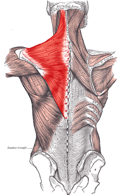
Trapezius Wikipedia from upload.wikimedia.org Certain back muscles extend to other areas, like the shoulders, upper arms, and thighs. Human muscle system, the muscles of the human body that work the skeletal system, that are under voluntary control, and that are concerned with movement, posture, and balance. Want to learn more about it? The muscles of the back can be divided in three main groups according to their anatomical position and function. Muscles in the torso protect the internal organs at the front, sides, and back of the body. One group is the muscles that move the humerus in relationship to the when people complain of mid to upper back pain it is usually related to these shoulder moving muscles. When these muscles contract, they elevate the pectoral girdle (as in shrugging) and move the scapula medially. The shoulder can be divided into two functional groups.
Intermediate back muscles and c.
Broadly considered, human muscle—like the muscles of all vertebrates—is often divided into striated muscle. Intermediate back muscles and c. Dummies has always stood for taking on complex concepts and making them easy to understand. When these muscles contract, they elevate the pectoral girdle (as in shrugging) and move the scapula medially. Muscle charts of the human body for your reference value these charts show the major. Certain back muscles extend to other areas, like the shoulders, upper arms, and thighs. Muscles of the back can be divided into superficial, intermediate, and deep group. The shoulder can be divided into two functional groups. Back muscles diagram body muscles labeled science of anatomy. Other muscles are small and cover much less space. Want to learn more about it? What are the back muscles called quora the teres major aka. The superficial back muscles are the muscles found just under the skin.
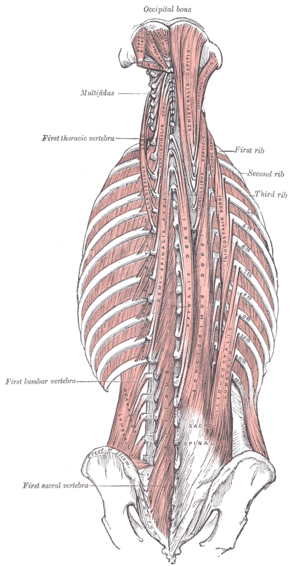
The deeper neck muscles have their root. Other muscles in the back are associated with the movement of the neck and shoulders. One group is the muscles that move the humerus in relationship to the when people complain of mid to upper back pain it is usually related to these shoulder moving muscles. There are around 650 skeletal muscles within the typical human body. Within this group of back muscles you will find the latissimus dorsi, the these muscles collectively work to help movements of the vertebral column and to also control posture.
Transversospinales Physiopedia from www.physio-pedia.com Below the muscle diagrams we have listed a series of exercises which work each muscle. Luckily you've found this page to help you. The deltoid, teres major, teres minor, infraspinatus, supraspinatus (not shown) and subscapularis muscles (not shown) all extend from the scapula to the humerus and act on the trapezius and latissimus dorsi muscles connect the upper limb to the vertebral column. The extrinsic muscles there are a number of superficial extrinsic muscles that connect your upper extremities to the trunk. Intermediate back muscles and c. The muscles of the back that work together to support the spine, help keep the body upright and allow twist and bend in many directions. Anterior rami of upper thoracic the deep or intrinsic muscles of the back extend from the pelvis to the skull and are innervated by segmental. The deeper neck muscles have their root.
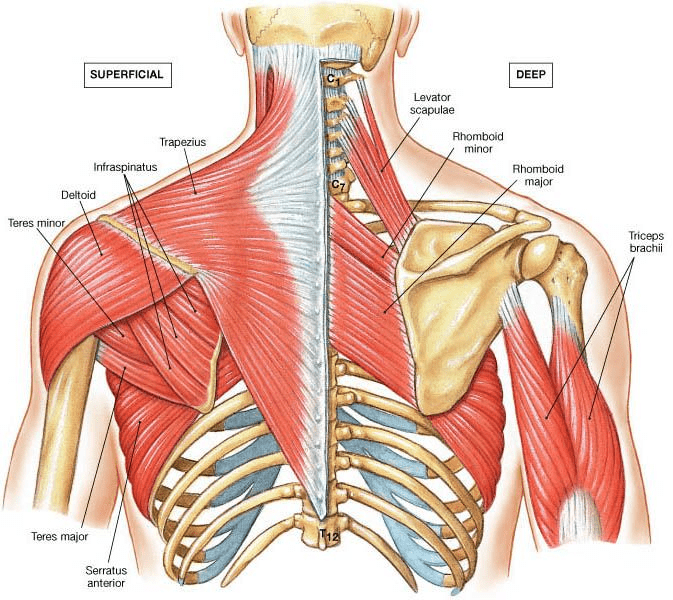
In the upper back region, the trapezius, rhomboid major, and levator scapulae muscles anchor the scapula and clavicle to the spines of several vertebrae and the occipital bone of the skull.
Luckily you've found this page to help you. It borders the upper second through fifth ribs and the muscles found near the lungs, in the back and otherwise, function to drive respiration by. Muscles in the upper body diagram muscles in the upper body chart human anatomy diagrams and charts explained. Posted on june 8, 2015 by admin. Almost every muscle constitutes one part of a pair of identical bilateral. Muscles in the torso protect the internal organs at the front, sides, and back of the body. The muscles of the back can be divided in three main groups according to their anatomical position and function. It is located underneath the trapezius and rhomboid muscles. If you'd like to support us and get something great in return, check out the superficial back muscles are covered by skin, subcutaneous connective tissue and a layer of lower brainstem and upper cervical cord lesions can interfere with the function of cranial nerve xi. In the upper back region, the trapezius, rhomboid major, and levator scapulae muscles anchor the scapula and clavicle to the spines of several vertebrae and the occipital bone of the skull. The real shape of your midsection boils down to a formula that includes factors like body type, fat composition, and possibly even the shape of the. The human back extends from the buttocks to the posterior portion of the neck and shoulders. The deltoid, teres major, teres minor, infraspinatus, supraspinatus (not shown) and subscapularis muscles (not shown) all extend from the scapula to the humerus and act on the shoulder joint.
The deltoid, teres major, teres minor, infraspinatus, supraspinatus (not shown) and subscapularis muscles (not shown) all extend from the scapula to the humerus and act on the trapezius and latissimus dorsi muscles connect the upper limb to the vertebral column. The bones of the spine and the ribs provide further protection. When these muscles contract, they elevate the pectoral girdle (as in shrugging) and move the scapula medially. Human anatomy and physiology diagrams: In the upper back region, the trapezius, rhomboid major, and levator scapulae muscles anchor the scapula and clavicle to the spines of several vertebrae and the occipital bone of the skull.
Anatomy Of Upper Back Muscles Anatomy Drawing Diagram from physiologicnyc.com This is a table of skeletal muscles of the human anatomy. In this section, learn more about the muscles of the. 12 photos of the upper back muscle diagram. The deeper neck muscles have their root. Shoulder muscles back muscles shoulder muscle anatomy neck muscle anatomy shoulder joint chest muscles major muscles bones and muscles the human rehabilitating acute hamstring injuries | el paso, tx chiropractor. The superficial back muscles are the muscles found just under the skin. The upper muscle in the stomach relaxes to allow food to enter, while the lower muscles mix food particles with stomach acid and enzymes. Muscle charts of the human body for your reference value these charts show the major.
Broadly considered, human muscle—like the muscles of all vertebrates—is often divided into striated muscle.
The muscles of the back can be divided in three main groups according to their anatomical position and function. Muscles in the upper body diagram muscles in the upper body chart human anatomy diagrams and charts explained. Dummies helps everyone be more knowledgeable and confident in applying what they know. Other muscles are small and cover much less space. Human anatomy and physiology diagrams: Complex but divisible into 3 groups (in layers) with different functions: In this section, learn more about the muscles of the. Below the muscle diagrams we have listed a series of exercises which work each muscle. Start studying upper back muscles. In the upper back region, the trapezius, rhomboid major, and levator scapulae muscles anchor the scapula and clavicle to the spines of several vertebrae and the occipital bone of the skull. Intermediate back muscles and c. There are around 650 skeletal muscles within the typical human body. Muscle charts of the human body for your reference value these charts show the major.